Tuesday, July 5, 2011

CASE : SPLENIC ARTERY PSEUDOANURISM BLEEDING Status TWICE COILING Status Chronic pancreatitis (alcoholic)^-www.drkeyurbhatt.in*

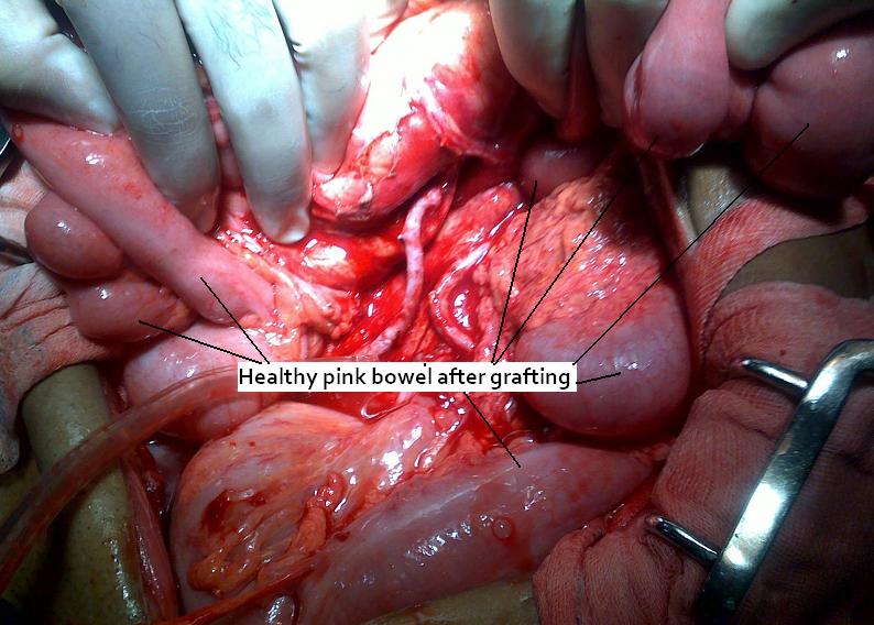
after 9 months of follow up Pt is doing well and gained 8 kg wt....no further complaints...


previous angio coiling, patent pancreatica
megna branch and partially occluded splenic artery


ligation of splenic artery at its origin before
arteria pancreatic magna branch


pseudoanurism in arteria pancreatica
magna teritory


Pancreatic Pseudoaneurysm: Treatment

  • Transarterial catheter angioembolization with or without endoscopic stent placement. 
o    Endovascular coil embolization has been used extensively in the treatment of visceral artery pseudoaneurysms.
o    Angioembolization is considered much less invasive than surgery. The procedure can be completed quickly and is comfortable for the patient. It also allows the performance of surgery under optimal conditions.
o    The interventional approach has a reported success rate of 67-100% over the past few years.6
o    Most authorities agree that embolization is appropriate when bleeding is diffuse or emanating from the pancreatic head, for unsuccessful surgery, or during postoperative bleeding.
o    Failure results from an inability to selectively catheterize the bleeding vessel or the misplacement or poor placement of embolization material.
o    In addition to rebleeding, complications of this procedure include rupture of the pseudoaneurysm during embolization, arterial perforation by the catheter, intestinal necrosis, and aortic thrombosis.


SURGICAL TREATMENT

The indications for emergent exploratory laparotomy include hemodynamic instability and failure of endovascular techniques to control active hemorrhage.

Basic surgical techniques for controlling hemorrhage from a pancreatic pseudoaneurysm include arterial ligation on both sides of the bleeding sites, pancreatic resection, and intracystic/extracystic multiple ligatures

Clinical features and management of splenic artery
pseudoaneurysm: Case series and cumulative
review of literature

(J Vasc Surg 2003;38:969-74.)

Deron J. Tessier, MD,a William M. Stone, MD,a Richard J. Fowl, MD,a Maher A. Abbas, MD,a James C.
Andrews, MD,b Thomas C. Bower, MD,c and Peter Gloviczki, MD,c Scottsdale, Ariz; and Rochester, Minn

Introduction: Splenic artery pseudoaneurysm is uncommon. We report our institution’s recent 18-year experience with
these aneurysms and review the literature.
Methods: We reviewed the records for 37 patients with visceral artery pseudoaneurysm evaluated at our institution from
1980 to 1998. From this group we found only 10 patients (27%) with splenic artery pseudoaneurysm. We also reviewed
147 cases of splenic artery pseudoaneurysm reported in the English literature.
Results: In this series of 10 patients, 5 were men. Mean age was 51.2 years (range, 35-78 years). Causes of aneurysm
included chronic pancreatitis in 4 patients, trauma in 2 patients, iatrogenic cause in 1 patient, and unknown cause in 3.
The most common symptom was bleeding in 7 patients and abdominal or flank pain in 5 patients; 2 patients had no
symptoms. Aneurysm diameter was known for four pseudoaneurysms, and ranged from 0.3 to 3 cm (mean, 1.7 cm).
Splenectomy and distal pancreatectomy were performed in 4 patients, splenectomy alone in 2 patients, endovascular
transcatheter embolization in 2 patients, and simple ligation in 1 patient. One patient with a ruptured pseudoaneurysm
died before any intervention could be performed; there were no postoperative deaths. Follow-up data were available for 7 patients, with a mean of 46.3 months (range, 4.5-120 months).

Conclusions: Splenic artery pseudoaneurysm is rare and usually is a complication of pancreatitis or trauma. Average aneurysm diameter in our series of 10 patients was smaller than previously reported (1.7 cm vs 5.0 cm). Although conservative management has produced excellent results in some reports, from our experience and the literature, we recommend repair of all splenic artery pseudoaneurysms. 


www.gisurgerysurat.com/

www.drkeyurbhatt.in/

www.sidshospital.com


Dr. Keyur Bhatt - Best Gastro Surgeon

Dr. Keyur Bhatt- Best GI Surgeon

Dr. Keyur Bhatt - Best Gastro Surgeon

Dr. Keyur Bhatt- Best GI Surgeon - Dr Keyur Bhatt - Best Gastro Surgeon

Dr. Keyur Bhatt- Best GI Surgeon - Dr Keyur Bhatt - Best Gastro Surgeon

                                   Dr Keyur Bhatt- Best GI Surgeon     


CASE: Abdominal angina with Total occlusion of all mesenteric vessels -> Ilea SMA & IMA Bypass grafting^-www.drkeyurbhatt.in*


after three months follow up patient gained 11 kg of wt.
and back to normal life working in fields...........


56 yrs male, chronic bidi smoker
had abdominal angina in 2008
h/o severe post parandial abdominal pain and wt loss of 25 kg in 2-3 months

Was diagnosed as chronic aortic dissection with atheromatous changes and occlusion of all mesenteric vessels..
Angiography of abdomen done in march 2008 (Mumbai) and SMA Was stented....

Pt recovered well after stenting and started gaining wt. & remained  asymptomatic for 1.5 yrs..
Again abdominal angina started and wt loss as well (32 kg)
Re angiography tried but stent could not be opened and other vessels were also completely occluded.

Referred for abdominal bypass.

CECT  ANGIO: Showed total occlusion of all mesenteric vessels:

Non visulization of SMA, IMA & CELIAC


SURGERY: Rt Ileo SMA (Superior mesenteric artery) and Left Ileo IMA (Inferior mesenteric artery) Bypass grafting with sehpanous vein. was done.(with team of  Myself,  Dr. Jaswantbhai Patel & Dr.Chetan Patel) and of course team of anaesthesia, radiology and intenciviest of Mahavir hospital.


AFTER GRAFTING:


POST SURGERY:

Patient started having regular meals after 3 months without pain and was discharged on POD- 5.


www.gisurgerysurat.com

www.sidshospital.com

You Tube : Dr Keyur Bhatt

www.gisurgerysurat.com/

www.drkeyurbhatt.in/

www.sidshospital.com


Dr. Keyur Bhatt - Best Gastro Surgeon

Dr. Keyur Bhatt- Best GI Surgeon

Dr. Keyur Bhatt - Best Gastro Surgeon

Dr. Keyur Bhatt- Best GI Surgeon - Dr Keyur Bhatt - Best Gastro Surgeon

Dr. Keyur Bhatt- Best GI Surgeon - Dr Keyur Bhatt - Best Gastro Surgeon

                                   Dr Keyur Bhatt- Best GI Surgeon     


Tuesday, June 28, 2011

Current status of minimally invasive necrosectomy for post-inflammatory pancreatic necrosis^-www.drkeyurbhatt.in*

REVIEW ARTICLE

Hepatobiliary Surgical Unit, Manchester Royal Infirmary, Manchester, UK


Abstract
Objective: This paper reviews current knowledge on minimally invasive pancreatic necrosectomy.
Background: Blunt (non-anatomical) debridement of necrotic tissue at laparotomy is the standard
method of treatment of infected post-inflammatory pancreatic necrosis. Recognition that laparotomy may
add to morbidity by increasing postoperative organ dysfunction has led to the development of alternative,
minimally invasive methods for debridement. This study reports the status of minimally invasive necrosectomy
by different approaches.

Methods: Searches of MEDLINE and EMBASE for the period 1996–2008 were undertaken. Only studies
with original data and information on outcome were included. This produced a final population of 28
studies reporting on 344 patients undergoing minimally invasive necrosectomy, with a median (range)
number of patients per study of nine (1–53). Procedures were categorized as retroperitoneal, endoscopic
or laparoscopic.

Results: A total of 141 patients underwent retroperitoneal necrosectomy, of whom 58 (41%) had
complications and 18 (13%) required laparotomy. There were 22 (16%) deaths. Overall, 157 patients
underwent endoscopic necrosectomy; major complications were reported in 31 (20%) and death in seven
(5%). Laparoscopic necrosectomy was carried out in 46 patients, of whom five (11%) required laparotomy
and three (7%) died.


Conclusions: Minimally invasive necrosectomy is technically feasible and a body of evidence now
suggests that acceptable outcomes can be achieved. There are no comparisons of results, either with
open surgery or among different minimally invasive techniques.

Received 31 October 2008; accepted 27 January 2009

www.gisurgerysurat.com

www.sidshospital.com

You Tube : Dr Keyur Bhatt

www.gisurgerysurat.com/

www.drkeyurbhatt.in/

www.sidshospital.com


Dr. Keyur Bhatt - Best Gastro Surgeon

Dr. Keyur Bhatt- Best GI Surgeon

Dr. Keyur Bhatt - Best Gastro Surgeon

Dr. Keyur Bhatt- Best GI Surgeon - Dr Keyur Bhatt - Best Gastro Surgeon

Dr. Keyur Bhatt- Best GI Surgeon - Dr Keyur Bhatt - Best Gastro Surgeon

                                   Dr Keyur Bhatt- Best GI Surgeon     


Recent advances in the surgical management of necrotizing pancreatitis^-www.drkeyurbhatt.in*


Department of Surgery, Meilahti Hospital, University of Helsinki, Helsinki, Finland
2005 Lippincott Williams & Wilkins.
1070-5295


Underdiagnosed and untreated, abdominal compartment syndrome is a potential contributing factor to the
development of early organ failure in patients with severe acute pancreatitis and warrants routine measurement of intra-abdominal pressure in patients treated for severe pancreatitis. The current estimate of the prevalence of intra-abdominal hypertension in severe acute pancreatitis is about 40%, with about 10% overall developing abdominal compartment syndrome, associated with increased hospital mortality rates. Early surgical decompression without exploring the pancreas further seems to be the most effective treatment. Primary fascial closure of the abdominal wall following abdominal decompression can be attempted, but in most cases the prolonged inflammatory process in the abdomen and the risk of recurrent abdominal compartment syndrome favors use of gradual closure or delayed reconstruction of the abdominal wall.
Summary
Recent studies confirm the overall validity of the established surgical principles for necrotizing pancreatitis:
delayed necrosectomy in patients with infected peripancreatic necrosis, mostly nonoperative management
of sterile necrosis, and delayed cholecystectomy in severe gallstone-associated pancreatitis. The role of abdominal compartment syndrome as an important contributing factor to early development of multiple organ failure and the potential benefit of surgical decompression are gaining support from recent reports and should be carefully assessed in future studies.


www.gisurgerysurat.com/

www.drkeyurbhatt.in/

www.sidshospital.com


Dr. Keyur Bhatt - Best Gastro Surgeon

Dr. Keyur Bhatt- Best GI Surgeon

Dr. Keyur Bhatt - Best Gastro Surgeon

Dr. Keyur Bhatt- Best GI Surgeon - Dr Keyur Bhatt - Best Gastro Surgeon

Dr. Keyur Bhatt- Best GI Surgeon - Dr Keyur Bhatt - Best Gastro Surgeon

                                   Dr Keyur Bhatt- Best GI Surgeon     


Sunday, June 19, 2011

CASE: Chronic pancreatitis - LPJ^-www.drkeyurbhatt.in*

26YRS female with pain in abdomen for 2 yrs
diagnosed as chronic pancreatitis

X RAY:

CECT;

Surgery: LPJ.


www.gisurgerysurat.com

www.sidshospital.com

You Tube : Dr Keyur Bhatt

www.gisurgerysurat.com/

www.drkeyurbhatt.in/

www.sidshospital.com


Dr. Keyur Bhatt - Best Gastro Surgeon

Dr. Keyur Bhatt- Best GI Surgeon

Dr. Keyur Bhatt - Best Gastro Surgeon

Dr. Keyur Bhatt- Best GI Surgeon - Dr Keyur Bhatt - Best Gastro Surgeon

Dr. Keyur Bhatt- Best GI Surgeon - Dr Keyur Bhatt - Best Gastro Surgeon

                                   Dr Keyur Bhatt- Best GI Surgeon