Wednesday, January 12, 2011

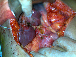
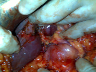
CASE: MASSIVE LOWER GI BLEEDING..With multiple Ileal ulcers...? Enteric ? Aspirin induced^-www.drkeyurbhatt.in*

55 yrs female with massive lower GI bleeding...with Hb 4.5 after 6 blood transfusions..
UGIE: was noraml..
LGIE: s/o few non bleeding ulcers in cecum...but ileum full of blood...

was kept conservatively and went into shock with NOR -ADR on 5 ml / hr..and 10 more transfusion was given ... and taken for mesantirc angio- which was inconclusive ---- I got a call.... decision of surgery was taken immediately and  Pt was explored....
and found to have hundreds of ileal ulcers in terminal 2 feets of ileum ....Intra of enteroscopy was done (by gastro physician, which helped us in defining proximal margin  free of ulcers)


 and along with ileal resection Rt hemicolectomy as done with I-T anastomosis....finally pt is walking today...passing normal stool...POD 5.

www.gisurgerysurat.com/

www.drkeyurbhatt.in/

www.sidshospital.com


Dr. Keyur Bhatt - Best Gastro Surgeon

Dr. Keyur Bhatt- Best GI Surgeon

Dr. Keyur Bhatt - Best Gastro Surgeon

Dr. Keyur Bhatt- Best GI Surgeon - Dr Keyur Bhatt - Best Gastro Surgeon

Dr. Keyur Bhatt- Best GI Surgeon - Dr Keyur Bhatt - Best Gastro Surgeon

                                   Dr Keyur Bhatt- Best GI Surgeon     


Sunday, January 9, 2011

CASE: Lower CBD stricture with multiple stones and Multiple Gall Stones..and Sigmoid MPD

65 yrs female with Obstructive jaundice...
USG s/o GSD + CBDS and lower CBD stricture...? CDC 1.
MRI / SVE  S/O lower CBD benign stricture and multiple stones...and Sigmoid MPD.(a congenital anomaly)
ERC failed...stenting done...


CEA / CA 19.9....and Brush Cytology was negative...

CDD-Choledecho Duodenostomy and Cholecystectomy was done...


completed CDD single layer interrupted vicryl

Thursday, January 6, 2011

CASE: SMV THROMBO EMBOLISM AND BOWEL GANGREEN^-www.drkeyurbhatt.in*

35 YRS male with severe agonizing pain in abdomen...
Past history: Operated for buccal malignancy few months ago..SOHND...on Chemotherapy and Radio Rx...On steriod...
CECT: S/O SMV thrombosis and gangrenous small bowel with air in wall of small bowel...
Pre Operatively on Inotrops and septic shock...

Anesthesia: Nasally intubated with bronchoscopy in view of inability to open mouth and SMF.
Explored: Total necrosis of whole jejunum...with peritonitis...
Surgery: Whole jejunum resected and Ileum anastomosed to DJ flexure..lavage and drainage...
post op remained on Venti for 4 days later converted to Tracheostomy...Gradually improved and came out of sepsis...supports weaned off....now taking oral diet and Ambulant.....Repeat CECT...revalled no leak and healthy bowel....on the line of discharge....


www.gisurgerysurat.com/

www.drkeyurbhatt.in/

www.sidshospital.com


Dr. Keyur Bhatt - Best Gastro Surgeon

Dr. Keyur Bhatt- Best GI Surgeon

Dr. Keyur Bhatt - Best Gastro Surgeon

Dr. Keyur Bhatt- Best GI Surgeon - Dr Keyur Bhatt - Best Gastro Surgeon

Dr. Keyur Bhatt- Best GI Surgeon - Dr Keyur Bhatt - Best Gastro Surgeon

                                   Dr Keyur Bhatt- Best GI Surgeon     


Wednesday, January 5, 2011

Isolated Pancreatic Tuberculosis^-www.drkeyurbhatt.in*

JOP. J Pancreas (Online) 2006; 7(2):205-210.
University of Pittsburgh School of Medicine. Pittsburgh, PA, USA
CASE REPORT
Context:  Pancreatic tuberculosis is an extremely rare clinical entity, despite the high prevalence of tuberculosis worldwide. The pancreas is protected from direct environmental exposure; therefore most cases of pancreatic tuberculosis arise from contiguous infection from peri-pancreatic lymph nodes or rarely from hematogenous
spread. Pancreatic tuberculosis can present as a cystic or solid pancreatic mass mimicking pancreatic malignancy. Diagnosing pancreatic tuberculosis is a clinical challenge and most cases are diagnosed after surgical exploration for presumed pancreatic cancer. Endoscopic ultrasound-guided fine needle aspiration (EUS-FNA) is being used more frequently for imaging and sampling of pancreatic lesions. Immediate cytopathologic examination of tissue sampled by EUS increases the diagnostic yield and is standard in many
institutions.

Isolated Pancreatic Tuberculosis
KUWAIT MEDICAL JOURNAL
Kuwait Medical Journal 2004, 36 (4):290-292
CASE REPORT
Tuberculosis of the pancreas is a clinical rarity and mimics pancreatic carcinoma both clinically and
radiologically. A 3 2 - y e a r-old Somali male patient presented with history of vague abdominal pain, weight loss, anorexia and jaundice. Radiological imaging showed gall stones, dilated common bile duct (CBD) and a hetrogenous pancreatic mass. Endoscopic retrograde cholangio pancreatography (ERCP) showed marked narrowing of the CBD with an impression of external compression. Cholecystectomy and  holedochoduodenostomy (CDD) were performed after frozen section histopathology revealed the mass to be tuberculosis. P reoperative diagnosis of pancreatic tuberculos is requires a high index of suspicion and usually its diagnosis is established after surgical tre a t m e n t . T h e response to antituberculosis treatment is very effective.

www.gisurgerysurat.com/

www.drkeyurbhatt.in/

www.sidshospital.com


Dr. Keyur Bhatt - Best Gastro Surgeon

Dr. Keyur Bhatt- Best GI Surgeon

Dr. Keyur Bhatt - Best Gastro Surgeon

Dr. Keyur Bhatt- Best GI Surgeon - Dr Keyur Bhatt - Best Gastro Surgeon

Dr. Keyur Bhatt- Best GI Surgeon - Dr Keyur Bhatt - Best Gastro Surgeon

                                   Dr Keyur Bhatt- Best GI Surgeon     


CASE: ? PRIMARY TUBERCULOSIS OF PANCREAS AND PERI PANCREATIC NODES^-www.drkeyurbhatt.in*

13 Yrs child with h/o pain in epigastric region for last 5 yrs..with back radiation. . . associated with nausea...anorexia....failure to thrive...
was ultimately recently diagnosed as chronic pancreatitis...with dilated duct...and bulky head....
ERCP tried but cannulation was failed....

Referred for surgical management....rest of the work up was completed and diagnosis of TROPICAL VARIETY OF CHRONIC  PANCREATITIS was made..

CECT:


MRI:


SURGERY: LATERAL PANCREATICO JEJUNOSTOMY
FINDINGS: Were Caseating node over head of pancreas...near gastro colic vein...and peri pancreatic few nodes with caseation milking out cheesy material...
MPD was 10 mm and full of cheesy pus......


drained and LPJ done....


www.gisurgerysurat.com/

www.drkeyurbhatt.in/

www.sidshospital.com


Dr. Keyur Bhatt - Best Gastro Surgeon

Dr. Keyur Bhatt- Best GI Surgeon

Dr. Keyur Bhatt - Best Gastro Surgeon

Dr. Keyur Bhatt- Best GI Surgeon - Dr Keyur Bhatt - Best Gastro Surgeon

Dr. Keyur Bhatt- Best GI Surgeon - Dr Keyur Bhatt - Best Gastro Surgeon

                                   Dr Keyur Bhatt- Best GI Surgeon